
Female Fertility Blood Panel Fertility Testing Meaning: AMH, Basal FSH, Estradiol, Thyroid & Prolactin Workups for Conception Planning
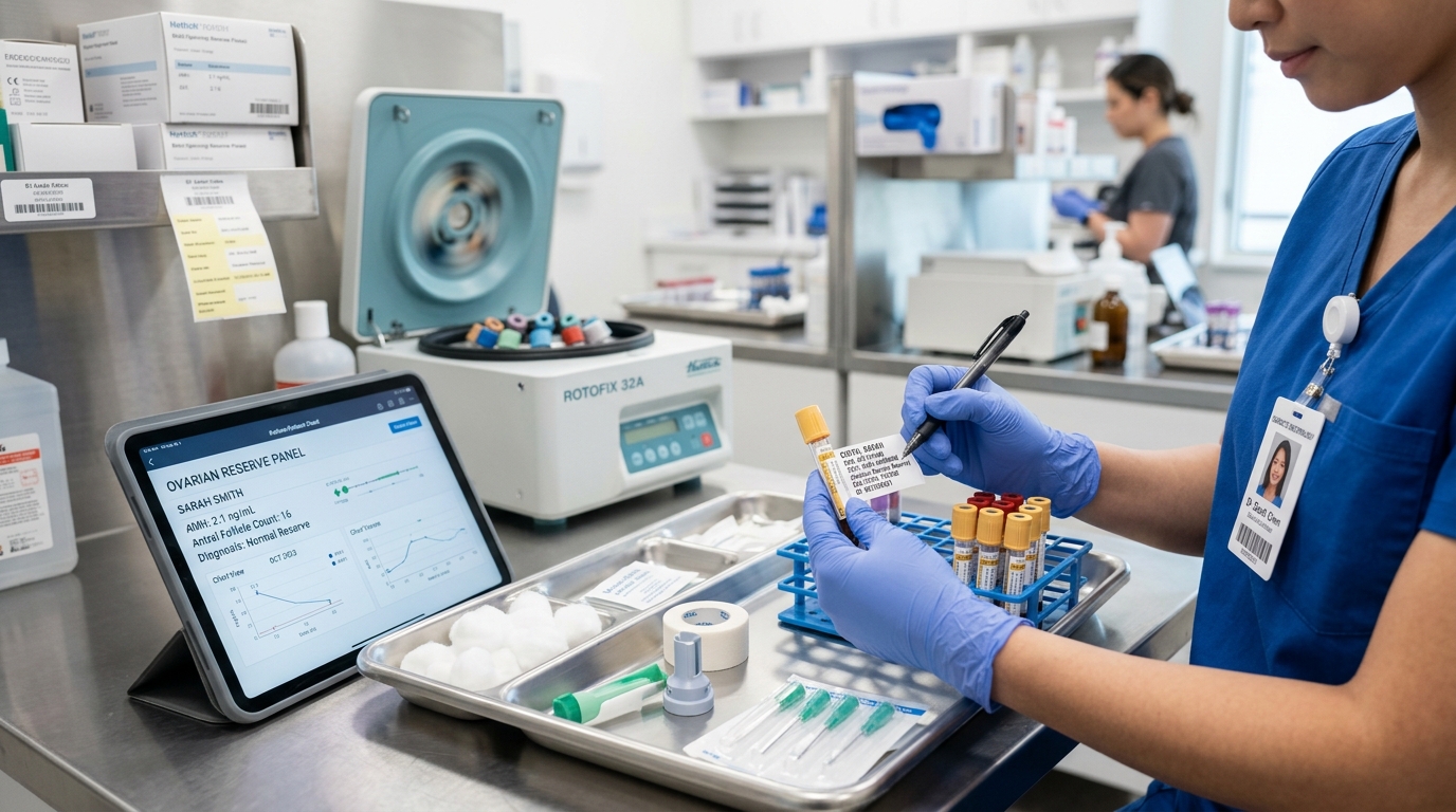
In modern fertility centers and women’s health clinics across Dubai, Abu Dhabi, and the wider UAE, proactive hormonal screening empowers couples to make informed family planning decisions. Sourcing validated professional fertility and diagnostic systems in Dubai ensures patients and physicians obtain reliable, precision-calibrated hormonal data.
The 5 Diagnostic Pillars of the Female Fertility Panel
To evaluate reproductive potential thoroughly, clinicians assemble laboratory biomarkers into five functional categories:
- Pillar 1: Ovarian Reserve (Egg Quantity): Measured by Anti-Müllerian Hormone (AMH) and Day 2–4 Basal FSH/Estradiol. Quantifies the remaining primordial follicle pool and predicts the number of oocytes that can be retrieved during IVF ovarian stimulation.
- Pillar 2: Ovulation Verification: Measured by Mid-Luteal Serum Progesterone (drawn 7 days prior to expected menses). Provides definitive biochemical confirmation that follicular rupture occurred and a functioning corpus luteum was established.
- Pillar 3: The Endocrine Modulators: Measured by Thyroid-Stimulating Hormone (TSH) and Serum Prolactin. The thyroid and pituitary glands act as metabolic master regulators; subtle subclinical abnormalities shut down GnRH pulsatility, producing luteal phase deficiency or anovulation.
- Pillar 4: Androgen & PCOS Profiling: Measured by Total/Free Testosterone, DHEA-S, and 17-Hydroxyprogesterone. Differentiates Polycystic Ovary Syndrome (PCOS) from non-classical congenital adrenal hyperplasia and androgen-secreting tumors.
- Pillar 5: Preconception Metabolic Health: Measured by Fasting Blood Glucose, HbA1c, 25-Hydroxy Vitamin D, and Rubella/Varicella Serology. Optimizes embryonic implantation rates and prevents early miscarriage and congenital teratogenicity.
Comprehensive Reference Range & Clinical Interpretation Matrix
Clinical practice guidelines from the American Society for Reproductive Medicine (ASRM) and the European Society of Human Reproduction and Embryology (ESHRE) outline target reference limits:
| Biomarker / Hormone | Timing of Blood Draw | Optimal Preconception Target Range | Clinical & Diagnostic Significance |
|---|---|---|---|
| Anti-Müllerian Hormone (AMH) | Any day of cycle (Cycle-independent) | 1.5 – 3.5 ng/mL (Normal reserve) | < 1.0 ng/mL = Diminished ovarian reserve; > 4.0 ng/mL = High reserve / PCOS morphology. Direct quantitative proxy of follicle count. |
| Basal Follicle-Stimulating Hormone (FSH) | Day 2, 3, or 4 of active menstrual flow | < 8.0 mIU/mL | Reflects pituitary drive to stimulate ovaries. Values > 10–12 indicate ovarian resistance and diminished reserve; > 25 indicates ovarian insufficiency. |
| Basal 17-Beta Estradiol (E2) | Day 2, 3, or 4 of active menstrual flow | 20 – 50 pg/mL | Values > 60–80 pg/mL indicate premature follicular development and artificially suppress (mask) high FSH into falsely normal ranges. |
| Thyroid-Stimulating Hormone (TSH) | Fasting morning sample | 0.5 – 2.5 mIU/L (Strict fertility window) | TSH > 2.5 mIU/L in women trying to conceive increases early miscarriage risk and impairs blastocyst implantation; treated with Levothyroxine. |
| Serum Prolactin | Mid-morning resting sample | < 20 ng/mL | Elevated prolactin disrupts GnRH pulsatility, producing short luteal phases, oligomenorrhea, or galactorrhea; warrants pituitary evaluation. |
| Mid-Luteal Serum Progesterone | Day 21 of 28-day cycle (or 7 days post-LH surge) | > 3.0 ng/mL (Confirms ovulation); > 10 ng/mL optimal | Confirms functional corpus luteum; values < 3.0 ng/mL indicate anovulatory cycle requiring ovulation induction (Letrozole). |
The Critical Distinction: Egg Quantity vs Egg Quality
A vital counseling rule in reproductive endocrinology is dispelling the myth that low AMH or high Day-3 FSH means a woman cannot conceive naturally:
- AMH and FSH Measure Quantity, Not Quality: Ovarian reserve testing predicts how many eggs remain and how many eggs can be retrieved during an IVF stimulation cycle. It does not measure whether those eggs are genetically normal.
- The Age Factor (Quality Benchmark): Oocyte chromosomal quality (aneuploidy rate) is determined overwhelmingly by chronological age, not AMH. A 28-year-old woman with a low AMH of 0.7 ng/mL has fewer eggs, but roughly 70% to 80% of her eggs are genetically normal, and her monthly spontaneous conception rate remains relatively high. Conversely, a 43-year-old woman with a high AMH of 3.0 ng/mL has plenty of eggs, but over 80% are chromosomally abnormal (aneuploid).
Pre-Analytical Phlebotomy Rules: Timing is Everything
Fertility blood panels are uniquely vulnerable to timing errors that generate false diagnoses:
- The Day 2–4 Rule: FSH, LH, and Estradiol must be drawn on Day 2, 3, or 4 of active menstrual bleeding. Drawing these labs on Day 7, 14, or 21 is completely uninterpretable and wastes healthcare resources.
- Resting Prolactin Protocol: Prolactin is a stress-sensitive hormone. Emotional stress, venipuncture anxiety, breast stimulation, and sexual intercourse in the preceding 24 hours cause transient “spikes.” If prolactin is mildly elevated, the patient should rest quietly in a chair for 30 minutes before a repeat blood draw.
- Fasting State: TSH, fasting glucose, insulin, and lipid panels must be drawn following an 8 to 12-hour overnight fast.
Frequently Asked Questions (FAQ)
Can a woman take a fertility blood panel while on birth control pills?
No. Combined oral contraceptive pills, hormonal IUDs, injections, and implants suppress the hypothalamic-pituitary-ovarian axis. Testing while on hormonal contraception will falsely show suppressed FSH, LH, Estradiol, and often an artificially reduced AMH (by 20%–30%). Patients should discontinue hormonal contraception for at least 2 to 3 natural cycles before undergoing definitive fertility testing.
Why is the fertility TSH target (0.5–2.5 mIU/L) stricter than the general lab normal range?
While standard laboratory reference ranges define normal adult TSH as up to 4.0 or 4.5 mIU/L, reproductive guidelines (ASRM and ATA) demonstrate that TSH levels between 2.5 and 4.0 in early pregnancy correlate with significantly higher miscarriage rates and lower live-birth rates. Maintaining preconception TSH < 2.5 ensures optimal thyroid reserve for early fetal neurodevelopment.
How does Vitamin D deficiency affect female fertility?
Vitamin D receptors are widely expressed in human ovarian granulosa cells and the endometrium. Clinical studies demonstrate that women with optimized Vitamin D levels (> 30 ng/mL) exhibit significantly higher clinical pregnancy and implantation rates during both natural conception and IVF compared to deficient women.
Authoritative Clinical References
- American Society for Reproductive Medicine (ASRM) – Diagnostic Evaluation of the Infertile Female: A Committee Opinion (2021)
- European Society of Human Reproduction and Embryology (ESHRE) – Assessment of Ovarian Reserve in Clinical Practice
- American Thyroid Association (ATA) – Guidelines for the Diagnosis and Management of Thyroid Disease During Pregnancy and Postpartum