
Pre-Analytical Error Prevention Checklist for UAE Labs: Phlebotomy Protocols, Specimen Rejection Criteria & ISO 15189 Quality Standards
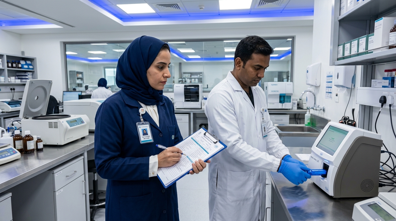
In diagnostic facilities and hospital networks throughout Dubai, Sharjah, and Abu Dhabi, establishing bulletproof pre-analytical workflows protects clinical reputations and ensures regulatory inspection excellence. Sourcing certified professional laboratory supplies and rapid diagnostic systems in the UAE guarantees healthcare facilities utilize vacuum tubes, lancets, and transport media manufactured to strict international tolerances.
The Anatomy of Pre-Analytical Error: The 70% Burden
In total testing process (TTP) science, diagnostic errors are categorized across three continuous phases:
- Pre-Analytical Phase (60% to 70% of all errors): Everything occurring prior to test analysis: test requisition, patient preparation (fasting/medication status), patient identification, phlebotomy technique, tube mixing, transport temperature, and centrifugation timing.
- Analytical Phase (< 10% to 15% of all errors): Analyzer mechanical pipetting, reagent calibration drift, optical sensor reading, and internal quality control execution. Due to high-level automation, analytical errors are now the rarest in modern laboratories.
- Post-Analytical Phase (15% to 20% of all errors): LIS transmission failures, delayed critical value phone calls, or physician misinterpretation of reference ranges.
The Master Pre-Analytical Prevention Checklist
Clinical and Laboratory Standards Institute (CLSI GP41 and GP44) and ISO 15189:2022 mandate adherence to a 10-point phlebotomy and accessioning checklist:
| Checklist Step | Standard Operating Procedure (SOP) Requirement | Clinical Failure Avoided | Accreditation Audit Standard |
|---|---|---|---|
| 1. Two-Identifier Verification | Active verbal confirmation of Full Name and Date of Birth; match with barcoded wristband/Emirates ID | Zero-tolerance for misidentified patients; eliminates transfusion and diagnostic tragedies. | JCI International Patient Safety Goal 1 (IPSG 1). |
| 2. Fasting & Medication Audit | Verbally confirm 8–12 hr fast for lipids/glucose; verify last dose of PPIs/antibiotics for stool/culture tests | Prevents lipemic specimen rejection and false-negative microbial tests. | ISO 15189:2022 Clause 7.2.4 (Pre-examination processes). |
| 3. Tourniquet < 60 Seconds | Release tourniquet within 1 minute of application; never allow fist pumping | Prevents hemoconcentration (falsely elevates potassium, calcium, total protein, and albumin). | CLSI GP41-A7 Phlebotomy Guidelines. |
| 4. Strict Order of Draw | Blood Cultures → Citrate (Blue) → Serum (Gold/Red) → Heparin (Green) → EDTA (Purple) → Fluoride (Gray) | Prevents additive cross-contamination (e.g., EDTA potassium carryover causing fatal pseudohyperkalemia). | CLSI GP41-A7 Mandatory Sequence. |
| 5. Proper Tube Inversion | Gently invert tubes 5 to 8 times immediately upon collection; never shake vigorously | Ensures anticoagulant mixing; prevents micro-clots in EDTA and hemolysis from mechanical shear. | CAP Laboratory General Checklist. |
| 6. Accurate Fill Volume (9:1 Ratio) | Citrate (Light Blue) tubes must be filled to ±10% of indicator line (9 parts blood to 1 part sodium citrate) | Underfilled citrate causes excess liquid anticoagulant, artificially prolonging PT/INR and aPTT coagulation times. | CLSI H21-A5 Coagulation Standards. |
| 7. Immediate Bedside Labeling | Affix barcode label onto the tube at the patient’s bedside; never pre-label tubes before draw | Pre-labeling tubes is the primary cause of sample swap errors in busy emergency and ICU wards. | DHA Health Regulation Standards. |
| 8. Centrifugation Window | Separate serum/plasma from red blood cells within 2 hours of collection (spin at 1,500–2,000 g × 10–15 min) | Delayed separation causes cellular glycolysis (falsely low glucose) and potassium leakage out of erythrocytes. | CLSI GP44-A4 Specimen Handling. |
| 9. Temperature-Controlled Transport | Validate insulated transport boxes with continuous digital temperature loggers (2°C–8°C for cold chain) | UAE ambient outdoor heat (> 40°C) rapidly degrades RNA, enzymes, and labile clotting factors. | DOH Abu Dhabi Specimen Transport SOP. |
| 10. Light Protection | Wrap specimens for bilirubin, vitamin A, B6, and carotene in aluminum foil or use amber tubes | Ambient fluorescent or sunlight exposure degrades photoreactive analytes within 30 minutes. | ISO 15189:2022 Clause 7.2.5. |
The 4 Major Specimen Rejection Criteria (HIL Interference)
Automated chemistry analyzers utilize optical spectrophotometric indices to quantify Hemolysis, Icterus, and Lipemia (HIL). Clear rejection protocols must be enforced:
- 1. In Vitro Hemolysis (Red/Pink Serum): Caused by small needle gauge (> 23G), pulling syringe plungers too hard, drawing through IV cannulas, or vigorous shaking. Lysis of erythrocytes releases intracellular contents: Potassium (K+), Lactate Dehydrogenase (LDH), AST, and Magnesium are falsely elevated. Troponin and coagulation assays are severely quenched. Samples exceeding manufacturer hemolysis index cutoffs must be rejected and redrawn.
- 2. Lipemia (Milky/Turbid Serum): Caused by high triglycerides (> 400 mg/dL) from non-fasting or metabolic disorders. Turbidity scatters light, interfering with photometric absorbance assays (electrolytes, hemoglobin, bilirubin).
- 3. Micro-Clots in Anticoagulated Blood (EDTA / Heparin): Inadequate initial tube inversion allows micro-thrombi to form. In complete blood counts (CBC), micro-clots falsely depress platelet counts (pseudothrombocytopenia) and clog automated hematology analyzer apertures.
- 4. Underfilled Sodium Citrate Coagulation Tubes: Sodium citrate binds calcium. If a tube is only half-filled, the relative excess of citrate chelates reagent calcium during the lab assay, yielding falsely elevated PT/INR and aPTT results that could lead to dangerous clinical over-anticoagulation interventions.
Middle East Climate Challenge: Transport Cold-Chain Auditing
In the UAE, summer ambient temperatures frequently exceed 45°C. Specimen transport from outpatient clinics or home collection to centralized reference laboratories represents a major pre-analytical vulnerability:
The Transport SOP: Phlebotomy transport couriers must utilize validated triple-packaging systems equipped with frozen gel packs and real-time digital temperature data loggers. Transport containers must maintain 2°C to 8°C throughout transit. If temperature data loggers indicate temperature excursions above 10°C for > 30 minutes, all molecular PCR, coagulation, and enzyme specimens must be rejected and recollected.
Frequently Asked Questions (FAQ)
Why must blood culture bottles always be drawn first in the order of draw?
Drawing non-sterile tubes first risks transferring skin commensal bacteria from the needle or tube stopper into subsequent bottles. Blood cultures must be drawn first to ensure the needle enters the sterile culture media without any carryover contamination.
Can a phlebotomist draw blood from an arm with an active intravenous (IV) infusion?
Strictly contraindicated. Drawing blood proximal to or through an active IV line introduces severe hemodilution from IV fluids (saline, dextrose), causing falsely low electrolyte, protein, and cell counts, alongside falsely elevated glucose or chloride. Blood should always be collected from the opposite arm. If unavoidable, the IV must be stopped for at least 2 minutes, and blood drawn distal to the cannula.
What is Pseudohyperkalemia, and how does pre-analytical error cause it?
Pseudohyperkalemia is an artificially elevated serum potassium level in a patient whose true physiological potassium is normal. Causes include: (1) drawing an EDTA (purple) tube before a chemistry (gold/green) tube, carrying potassium EDTA into the chemistry tube, (2) prolonged tourniquet application with fist clenching, (3) mechanical hemolysis, and (4) delayed centrifugation allowing potassium to leak out of red blood cells.
Authoritative Clinical References
- International Organization for Standardization – ISO 15189:2022 Medical Laboratories Requirements for Quality and Competence
- Clinical and Laboratory Standards Institute (CLSI) – GP41: Collection of Diagnostic Venous Blood Specimens
- Dubai Health Authority (DHA) – Diagnostic Laboratory Services Standards and Regulations