
What Does H. pylori Antibody Rapid Testing Detect—and What Can It Miss?
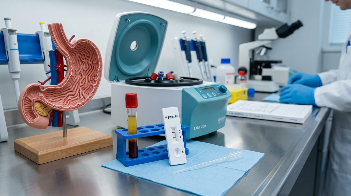
Epigastric burning, postprandial fullness, and persistent dyspepsia are among the most frequent gastrointestinal complaints evaluated by primary care physicians and gastroenterologists across the UAE. In community clinics and diagnostic screening centers, point-of-care rapid testing using gastrointestinal rapid diagnostic kits provides actionable results within 10 to 15 minutes. However, clinicians must distinguish when an antibody serology test is appropriate versus when an active infection assay—such as the H. pylori stool antigen rapid test or a broader food intolerance 286 panel for chronic digestive distress—is necessary to guide definitive eradication therapy.
Biological Target: Host Humoral IgG and IgM Immunoglobulins
Unlike stool antigen tests or Urea Breath Tests (UBT) that directly measure the physical bacterium or its metabolic urease enzyme activity, an H. pylori antibody rapid test detects the host immune response:
- Circulating Serum Anti-H. pylori IgG: When H. pylori colonizes the gastric epithelium, it provokes a vigorous systemic humoral response. High-affinity Immunoglobulin G (IgG) antibodies against surface antigens (CagA, VacA, Urease subunits) appear in peripheral blood within 3 to 4 weeks of colonization.
- Lateral Flow Detection: A whole blood, serum, or plasma sample is applied to the sample well. Recombinant H. pylori antigens immobilized on the nitrocellulose membrane capture host IgG antibodies, creating a visible pink-to-red colloidal gold test band.
Diagnostic Comparison: Antibody vs Stool Antigen vs Breath Test
Gastroenterology consensus guidelines (such as the Maastricht VI European Consensus and American College of Gastroenterology) classify H. pylori diagnostics into active vs non-active modalities:
| Diagnostic Methodology | What It Detects | Confirms Active Infection? | Useful for Post-Treatment Eradication? | Effect of PPIs / Antibiotics |
|---|---|---|---|---|
| H. pylori Blood Antibody Rapid Test | Host serum IgG antibodies | No (detects past or present exposure) | Strictly No (antibodies persist for years) | Unaffected (reliable even if taking PPIs) |
| H. pylori Stool Antigen Test (SAT) | Physical bacterial proteins in feces | Yes (indicates living colonization) | Yes (test 4–6 weeks post-antibiotics) | Must stop PPIs for 2 weeks & antibiotics for 4 weeks |
| ¹³C / ¹⁴C Urea Breath Test (UBT) | Bacterial urease enzyme activity | Yes (gold standard non-invasive) | Yes (test 4–6 weeks post-antibiotics) | Must stop PPIs for 2 weeks & antibiotics for 4 weeks |
| Endoscopic Biopsy (CLO / Histology) | Mucosal histology & rapid urease | Yes (invasive gold standard) | Used during follow-up endoscopy for ulcers | Direct tissue visualization |
What H. pylori Antibody Rapid Testing Can Miss: Diagnostic Limitations
Medical providers should recognize four distinct failure modes of serological testing:
1. Inability to Confirm Eradication
If a patient completed a 14-day quadruple therapy regimen (bismuth, metronidazole, tetracycline, PPI) six months ago and presents with recurrent dyspepsia, an antibody test will remain positive. Relying on serology in this scenario leads to inappropriate, repetitive antibiotic prescribing.
2. The Early Infection Window (Seroconversion Delay)
In acute primary infection (rarely captured in adults, but possible during outbreaks), it takes 2 to 4 weeks for serum IgG titers to cross the analytical detection threshold, leading to potential false-negative results in early acute gastritis.
3. Low Positive Predictive Value in Low-Prevalence Populations
In regions or demographics where H. pylori background prevalence has fallen below 20%, the Positive Predictive Value (PPV) of serology drops significantly, increasing the proportion of false positives relative to true active cases.
Clinical Decision Workflow for Suspected H. pylori
To ensure rational diagnostic stewardship:
- Treatment-Naïve Patient: In a young patient (< 50 years) with dyspepsia who has never been treated for H. pylori, a positive rapid antibody test can support empirical eradication therapy under a “test-and-treat” strategy.
- Previously Treated Patient: If the patient has a history of prior H. pylori treatment, NEVER order serology. Discontinue PPIs for 14 days and perform a Stool Antigen Test or Urea Breath Test.
- Alarm Symptoms (Red Flags): In patients over 50 or presenting with dysphagia (difficulty swallowing), unexplained weight loss, persistent vomiting, palpable epigastric mass, or gastrointestinal bleeding, bypass all non-invasive rapid tests and refer immediately for upper gastrointestinal endoscopy (EGD) with gastric biopsy.
Peer-Reviewed Clinical References
- Malfertheiner, P., et al. Management of Helicobacter pylori infection: the Maastricht VI/Florence consensus report. Gut, 2022; 71(9): 1724-1762.
- Chey, W. D., et al. ACG Clinical Guideline: Treatment of Helicobacter pylori Infection. The American Journal of Gastroenterology, 2017; 112(2): 212-239.
- Wang, Y. K., et al. Diagnosis of Helicobacter pylori infection: Current options and developments. World Journal of Gastroenterology, 2015; 21(40): 11221-11235.